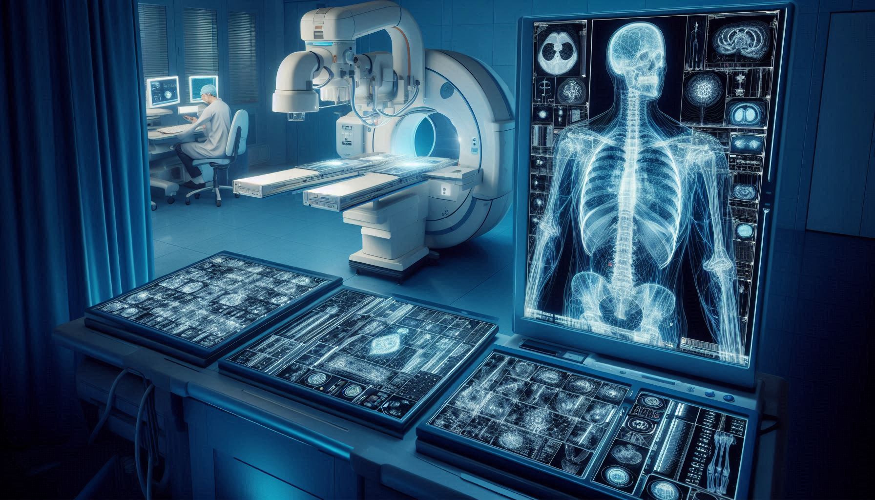
X-rays are a crucial diagnostic tool in modern medicine, helping doctors see inside the body to diagnose and monitor various conditions. But what technologies make X-ray imaging possible, and how have they evolved over time? This article delves into the key technologies and principles that drive the world of X-ray imaging.
1. Introduction to X-Ray Technology
X-rays are a form of electromagnetic radiation, similar to visible light but with much higher energy. This energy allows X-rays to pass through most objects, including the human body, making them ideal for medical imaging. The X-ray beam’s interaction with tissues and bones creates images that help physicians diagnose fractures, tumors, and other internal issues.
2. Basic Principles of X-Ray Imaging
2.1 How X-Rays Work
When X-rays pass through the body, they are absorbed at different rates by different tissues. Dense materials like bones absorb more X-rays and appear white on the resulting image, while softer tissues absorb fewer X-rays and appear in shades of gray.
2.2 The Role of Radiographic Film
Traditionally, X-rays were captured on radiographic film, similar to photography. Today, many facilities have moved on to digital sensors, which capture X-ray images electronically for immediate viewing and analysis.
3. Technologies Behind X-Ray Machines
Several core technologies contribute to the effectiveness of X-ray imaging:
3.1 X-Ray Tubes
The heart of any X-ray machine is the X-ray tube. This device generates X-rays by directing a stream of electrons at a metal target, typically made of tungsten. When the electrons collide with the metal, they decelerate rapidly, producing X-rays as a byproduct.
3.2 Detectors and Imaging Plates
In modern digital X-ray machines, detectors capture X-rays after they pass through the body. These detectors come in two main types:
- Direct Digital Radiography (DR): Uses flat-panel detectors to capture X-ray images directly and display them on a monitor almost instantly.
- Computed Radiography (CR): Uses imaging plates coated with phosphors that store the energy from X-rays. The plate is then scanned by a machine that reads and digitizes the image.
4. Advances in X-Ray Technology
Recent technological advances have significantly improved the quality, safety, and applications of X-ray imaging:
4.1 Digital X-Ray Systems
Digital systems offer higher resolution and faster results than traditional film-based systems. They reduce the need for repeated exposures, thus lowering the patient’s exposure to radiation.
4.2 3D X-Ray Imaging (Cone Beam CT)
Cone Beam Computed Tomography (CBCT) provides three-dimensional X-ray images by rotating the X-ray source and detector around the patient. This type of imaging is especially useful in dental and orthopedic applications, offering detailed views that traditional 2D X-rays cannot.
4.3 Portable X-Ray Machines
Technological miniaturization has led to the development of portable X-ray machines. These compact devices are invaluable in emergency settings, remote locations, or when patients cannot be moved to a conventional X-ray room.
5. Safety Technologies in X-Ray Systems
Despite their diagnostic power, X-rays expose patients and technicians to radiation. To minimize risks, several safety technologies are built into modern X-ray machines:
5.1 Lead Shielding
Lead aprons and shields are essential for protecting parts of the body that do not need to be imaged. Lead’s high density makes it excellent for blocking X-rays and minimizing exposure.
5.2 Collimators
A collimator narrows the X-ray beam to the specific area being imaged, reducing radiation exposure to surrounding tissues.
5.3 Automated Exposure Control (AEC)
This technology automatically adjusts the X-ray machine’s settings based on the patient’s size and the density of the targeted area, ensuring optimal image quality with minimal radiation.
6. Specialized X-Ray Technologies
X-ray technology has diversified to cater to different medical needs:
6.1 Fluoroscopy
Fluoroscopy provides real-time moving images of internal structures, which is useful for guiding certain procedures like catheter insertions or joint examinations.
6.2 Mammography
Mammography is a specialized form of X-ray imaging used to detect breast cancer. These machines are designed to provide high-resolution images of breast tissue for early detection.
6.3 Dual-Energy X-Ray Absorptiometry (DEXA)
DEXA scans measure bone density to assess conditions like osteoporosis. This technology uses two X-ray beams at different energy levels to determine bone density and body composition.
7. X-Ray Technology Beyond Medicine
While most commonly associated with healthcare, X-ray technology is used in other fields as well:
- Security: Airports use X-ray scanners to inspect luggage and ensure safety.
- Industrial Applications: Engineers use X-rays to check the structural integrity of machinery and materials.
8. Future Developments in X-Ray Technology
The future of X-ray technology looks promising, with ongoing advancements aimed at improving image quality and reducing radiation exposure. Innovations in AI integration are enabling faster and more accurate image analysis, while new materials and methods are being explored for even more precise imaging.
Conclusion
X-ray technology has come a long way since its discovery in 1895. From simple 2D film-based imaging to advanced 3D digital scans, it continues to be an invaluable tool for diagnosing and monitoring medical conditions. Understanding the technology behind X-ray imaging not only highlights its capabilities but also underscores the importance of safety measures that keep both patients and medical professionals protected.


